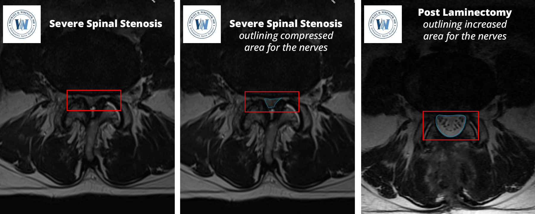
As we age, the lumbar (lower) spine sometimes develops a narrowing of the spinal canal due to wear and tear. This is caused by a combination of collapsed discs, overgrown bone spurs, and thickened ligament. The narrowing of the canal can lead to compression of the nerves that run down the center of the canal; this condition is called lumbar spinal stenosis.
Patients with spinal stenosis may experience leg pain and/or back pain from the compressed nerves. Symptoms are usually worse with activity such as standing or walking and improved with rest. When someone has symptoms from spinal stenosis, he or she should consult with a spine surgeon.
Lumbar laminectomy is a surgical procedure performed after conservative treatments including medication, injections, or physical therapy have failed. During a lumbar laminectomy, the pressure is taken off the compressed nerves – also known as a “decompression” procedure. A laminectomy may be necessary at only one level, or sometimes at multiple levels, depending on the patient’s symptoms and imaging.
Benefits of Lumbar Laminectomy
- Relief of activity-related back pain caused by the spinal stenosis
- When the spinal canal is narrowed (spinal stenosis) and pressure is placed on the nerves, patients may experience activity-related back pain. An example of this is pain with standing and walking around, and pain improves after sitting or lying down. By taking the pressure off the compressed nerves, patients may experience improvement in their back pain.
- Relief of leg pain, weakness, or numbness
- When there is compression of their nerves in the lumbar spine, the person may experience “leg symptoms”. These symptoms present in a predictable distribution, depending on which nerves are involved. Possible symptoms include weakness, numbness, tingling, or shooting pain down the legs. By taking the pressure off the affected nerves, dramatic improvement in the leg pain (or other symptoms) can be achieved.
- Increase activity tolerance
- Lumbar spinal stenosis often limits the distance one can walk or the amount of time one can participate in activity due to pain or other symptoms caused by the compression of the nerves. By taking the pressure off the compressed nerves, patients often notice a dramatic improvement in their ability to walk or stand for longer periods of time.
Patients who would benefit from a Lumbar Laminectomy
- Patients who have diagnosed spinal stenosis on an MRI or CT myelogram who have failed conservative treatment. Surgical treatment of spinal stenosis should only be performed when a patient has failed conservative care and quality of life is affected by the spinal stenosis.
- Patients who underwent spinal steroid injections that resulted in short term but excellent relief of symptoms are great candidates for a lumbar laminectomy.
- Prior to proceeding with a lumbar laminectomy, other causes of symptoms should be ruled out. Sometimes patients who have blood flow problems in the legs can mimic symptoms of spinal stenosis.
Pre and Post Laminectomy Images

Positioning: The patient is positioned prone (face down) and padding is placed to protect the face and bony areas. General anesthesia is used, meaning the patient will completely asleep for this surgery.
Incision: A vertical incision is made over the affected level(s) of the lower back.
Exposure: The muscles of the spine are carefully separated from the bone to allow access to the spine.
Confirmation of surgical level: X-ray is used to confirm the appropriate surgical level.
Laminectomy: This process involves removing the “lamina” of the bone. This can be done in its entirety, or only on one side. The decision to do a laminectomy vs laminotomy is usually made prior to surgery and depends on the patient’s symptoms and MRI. When only one side is removed, it is called a “laminotomy”. “-ectomy” means removal, while “-otomy” means to make a hole. The bone is carefully removed by using a burr, which is similar to a dremel tool.
Decompression: After the laminectomy, the next step is to expose the thecal sac which contains the spinal nerves. The ligament that covers the spinal nerves is removed. Once the spinal nerves are exposed, the next step is removing the culprit causing compression on the nerves. This usually involves removing overgrown bone spurs, parts of degenerated disc, and other non-essential structures to make sure all the pressure is off the nerves.
Incision closure: The incision is closed with absorbable suture and a dressing is placed.
Risks and complications associated with Lumbar Laminectomy
Although risks associated with lumbar laminectomy are rare, patients undergoing this surgery should be aware of the risks involved and discuss with Dr. Wheeler prior to surgery:
- Dural tear – sometimes there is a small tear in the outer tissue layer that contains the nerves, called the dura. If there is a tear, leakage of cerebral spinal fluid may occur. Most tears are small and repaired at the time of surgery without any consequence.
- Epidural hematoma – this rare complication can occur when a blood clot forms within the surgical bed after surgery, putting pressure on the nerves.
- Nerve damage
- Pain, bleeding, infection – which are risks of any surgery
Recovery
- Depending on the patient’s health and number of levels performed, the patient may discharge home the same day of surgery.
- A back brace is usually given to the patients to provide them with support during the recovery period.
- Complete recovery from surgery varies, but patients can usually return to normal activities 6-12 weeks after surgery.
- Patients can return to work as early as 1-2 weeks postoperatively depending on job responsibilities.


